
Brain tumours are abnormal growth of brain cells and can be benign or malignant (cancerous). In a broad sense, brain tumours can be primary or metastatic (from cancer of other parts of the body). Such tumours clinically manifest with various symptoms depending on their location, size, and histological variety. Common presentations of brain tumours are headaches, vomiting, and seizures, or epilepsy. Some may have focal neurological deficits like weakness of limbs, memory impairment, speech problems, restricted vision, gait disturbances etc.
The incidence of brain tumours is increasing and is 5 to 10 per 100,000 in the Indian population. They represent a substantial cause of morbidity and mortality and an overall decrease in productivity. The gradual increase in the incidence of brain tumours in young populations has also led to a significant financial burden on families. Early diagnosis and management has a better outcome in patients with brain tumours.
What are the causes of brain tumours?
The risk factors associated with brain tumours are ionising radiation, environmental factors, and mutations of genes controlling cell growth. Various hereditary conditions like neurofibromatosis and tuberous sclerosis are also associated with higher incidences of brain tumours.
What are the types of brain tumours?
The common primary brain tumours are gliomas, meningiomas, pituitary tumours and nerve sheath tumors. Gliomas are the most common intraparenchymal brain tumours and can be of low grade or highly malignant high grade. Secondary brain tumours or metastatic brain tumours originate from cancer in other parts of the body and spread to the brain through the blood or lymphatics. Cancers from lungs, breast, kidney, prostate, and melanoma commonly spread to the brain. The incidence of metastatic brain tumours is higher than primary tumors.
How are brain tumours diagnosed?
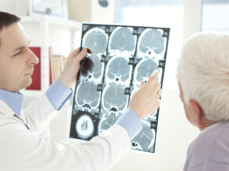
Brain tumours can be diagnosed on the basis of clinical symptoms. Some specific symptoms like early morning headache that subsides with vomiting, repeated seizures involving a particular area of the body (like hand, face, leg etc.), visual field restriction (narrowing of the visual field, being unable to see one half of the visual field) are characteristics of brain tumors. However, the diagnosis is confirmed with radiological evaluation. A CT scan of the head can grossly show the brain tumor. Additionally, various sequences of MRI provide details about the nature of tumour, like low grade or high grade. However, the exact nature of cellular origin and biological characteristics can be detailed by histopathological study of tumour tissue obtained from biopsy or surgery. This is also important in grading the tumour into the low grade or high grade variety.
What are the treatment options available for brain tumours?
There are several treatment options for brain tumors, including surgery, radiotherapy, chemotherapy, and gamma knife radiosurgery. Biopsy of the tumour may be considered for smaller intraparenchymal brain tumors. The MRI guided streotactic brain tumour biopsy provides accurate localisation of the brain tumour as well as tissue diagnosis or histopathological characterization of the brain tumour. Then, depending on the grade of tumour further therapy like radiation or chemotherapy can be planned for such patients. However, complete surgical excision is the best treatment for brain tumours and has a better long-term onco-functional outcome.
What are the risks involved in brain tumour surgery and how can they be reduced?
As the brain is the controller of entire body function, any additional injury to the nearby brain area during tumour surgery may result in functional impairment. Therefore, the brain tumour has to be resected out with minimal handling of the adjacent brain tissue. Various functional imaging techniques like functional MRI and diffusion tensor imaging are used to detect the functional brain areas around brain tumors. Again, such areas can be stimulated and preserved with the use of direct electrical stimulation in patients operated in awake conditions. Further use of intraoperative ultrasound also reduces the risk of injury to normal brain areas. Use of these advanced modalities allows better tumour removal even if the tumour is located in eloquent areas like insular glioma, brain stem glioma. A variety of brain tumour surgeries like glioma, vestibular schwannoma are also being used to minimise functional deficits after surgery.
Minimal invasive surgery for brain tumours like endonasal endoscopic procedures are now the standard of care for operating pituitary tumors.
Even large brain tumours located in the pituitary region can be operated safely through the nose using an endoscope.
What is gammaknife radiosurgery and which brain tumours are managed with this?
Gammaknife radiosurgery is a form of radiation therapy where radiation is precisely focused on the brain tumours with minimal effect on surrounding brain areas. Commonly, small, benign brain tumours like meningioma, vestibular schwannoma, cavernous hemangioma, cavernoma, etc. are treated with gamma knife radiosurgery. This has the ability to safeguard important functional brain areas like the cochlea (hearing apparatus), optic nerve (visual apparatus), and brain stem while giving an adequate dose to the brain tumour targeted for therapy. Apart from this gammaknife radiosurgery provides excellent results in patients with brain arteriovenous malformation.
What is the prognosis of a brain tumour?
The outcome of a brain tumour depends on the type (low grade vs. high grade), location, extent of tumour removal, and the patient’s clinical condition. Good tumour removal always has a better outcome in patients with low grade tumors. Therefore, maximal tumour removal is always attempted in these cases. However, high grade tumours like glioblastoma have a poor prognosis even after complete tumour removal and post-operative radiotherapy and chemotherapy. Apart from this, certain genetic mutations like TP53, IDH1/2, ATRX, 1p19q, etc also affect the prognosis.
Who should be screened for
brain tumours?
Patients who have already been diagnosed with cancer of the lungs, breast, kidney, or prostate and develop symptoms such as headache, vomiting, or seizures should be evaluated for a brain tumor. Any metastatic lesion can be detected using a CT scan or an MRI of the brain. People with a family history of cancer or previously diagnosed with hereditary syndromes like neurofibromatosis, tuberous sclerosis, Von Hippel-Lindau disease, and multiple endocrine neoplasia are prone to developing brain tumours and should be screened regularly.
Dr Sushant Kumar Sahoo is an Associate professor at Department of Neurosurgery and Gammaknife, Radiosurgery at PGIMER, Chandigarh.